Look for deep disease
Assess areas such as the uterosacral ligaments, pelvic sidewalls, bowel and bladder when included in the dedicated protocol.
 LONGEVITYPlan your visit
 SIMONMED LONGEVITY · HEALTHCHECK 2.2
Pelvic pain deserves
a deeper look.
A dedicated pelvic MRI can help map suspected deep endometriosis and related conditions, giving you and your care team more information for the next decision.

WHAT THIS EXAM ADDS
Endometriosis occurs when tissue similar to the lining of the uterus grows outside it. It can be associated with painful periods, persistent pelvic pain, pain with sex, bowel or bladder symptoms and fertility concerns. MRI can add detail when symptoms and earlier testing leave questions.[1]
Assess areas such as the uterosacral ligaments, pelvic sidewalls, bowel and bladder when included in the dedicated protocol.
Identify ovarian endometriomas and assess adenomyosis, a related condition involving the uterine muscle.
Help your gynecologist understand the location and extent of visible disease, including when surgical planning may require more than one specialist.
THE SCIENCE, EXPLAINED
The 2025 European Society of Urogenital Radiology consensus supports MRI when ultrasound is inconclusive or negative in a person with symptoms, and for selected treatment or surgical planning.[1]
Dedicated sequences and an experienced reader matter. A general pelvic MRI and an endometriosis-focused interpretation are not necessarily interchangeable.
Small or superficial endometriosis can be invisible on MRI. A negative scan does not exclude the condition; persistent symptoms deserve continued gynecologic care.
FOCUSED IMAGING
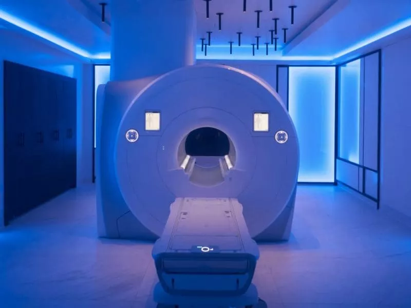
Modern endometriosis protocols use complementary MRI views to assess pelvic anatomy, blood products and signs of deep disease. IV contrast is not routinely required for typical endometriosis mapping, although selected findings may need a different protocol.[2]
This is a focused assessment for people with suspected disease or relevant symptoms, not a proven population-screening test for everyone without symptoms. Your clinician can help decide whether ultrasound, MRI or another evaluation is the best next step.
YOUR EXAM
Share symptoms, menstrual and surgical history, prior imaging and any fertility priorities. Follow the center’s preparation instructions.
Lie still for a dedicated pelvic MRI. The team explains positioning and confirms whether any special preparation or contrast is needed.
Review the report with your gynecologist. Imaging may guide treatment planning, but does not determine treatment on its own.
GOOD TO KNOW
No. Pelvic pain has many possible causes. MRI can add useful information, but your history, examination and other tests remain important.
It can help avoid uncertainty and improve planning, but it does not replace every diagnostic procedure. Your gynecologist will explain whether further evaluation is needed.
It requires additional imaging time and may be scheduled separately. Confirm timing and preparation with the concierge.
GO FURTHER, THOUGHTFULLY
CONCIERGE SIMONMED LONGEVITY SCHEDULING
xxx xxx xxxx
Locations nationwide · Exam availability varies by location.
Bundled with your $399 HealthCheck 2.0. Add-ons require clinical suitability and may need more time or a separate appointment.
Plan your visit ↗THE EVIDENCE BEHIND THE EXPLANATION
Sources reviewed September 19, 2026. Study populations and limitations are described above. Educational information complements individualized medical care.
SIMONMED LONGEVITY
Advanced Testing. Results. Plan.
Start with HealthCheck 2.0